Parts of digestive system:
- Mouth: Mouth is also called Oral cavity or buccal cavity. It is formed by cheeks, hard palate, soft palate and tongue.
- Cheeks form lateral walls of mouth and are covered by skin from outside and mucous membrane from inside.
- Hard palate forms anterior portion of roof of mouth. Hard palate is made up of palatine and maxillae bones covered with mucous membrane. Hard palate forms bony partition between oral and nasal cavity.
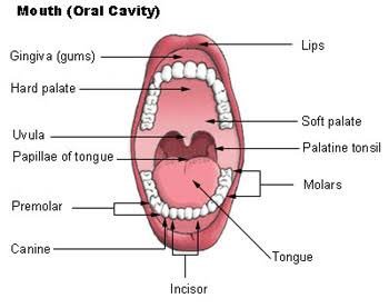
- Soft palate forms posterior portion of roof of mouth. It forms partition between oropharynx and nasopharynx. Soft palate is also covered with mucous membrane.
- Uvula is small muscular process hanging from soft palate. It prevents entrance of swallowed food and liquid into nasal cavity.
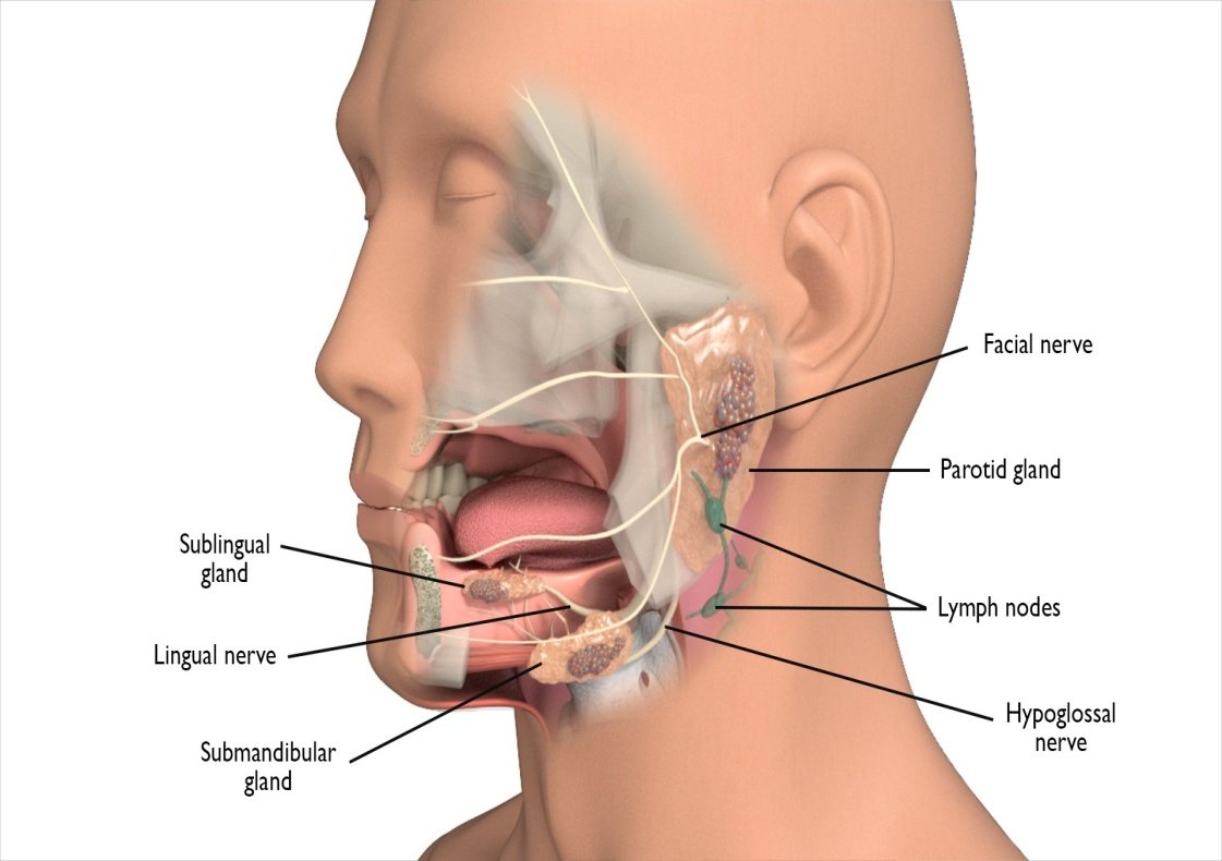
- Salivary glands: A salivary gland is a gland that releases a secretion called saliva into the oral cavity. Saliva is secreted to keep the mucous membranes of the mouth and pharynx moist and to cleanse the mouth and teeth. When food enters the mouth, secretion of saliva increases, and it lubricates, dissolves and begins the chemical breakdown of the food. There are three pairs of major salivary glands which secrete saliva:
- The parotid glands: (par = near; ot = ear) are located near ears. These secretes saliva into the oral cavity via a parotid duct that open into the vestibule opposite the second maxillary (upper) molar tooth.
- The sub-mandibular glands: (sub = below, mandible = lower jaw bone) are found in the floor of the mouth, below lower jaw. Their ducts open into the oral cavity lateral to the lingual frenulum.
- The sublingual glands: (sub = below, lingual = tongue) are beneath the tongue and superior to the sub-mandibular glands. Their ducts open into the floor of the mouth. Several minor glands are also present in cheeks, palates, tongue and lips etc. which produce small amount of saliva. Process of secretion of saliva is called salivation.
Composition and Functions of Saliva
Saliva is a vital fluid produced by the salivary glands in the mouth. It plays several important roles in digestion and oral health. The composition of saliva includes:
Composition of Saliva:
- Chemically saliva consists of 99.5% water and 0.5% of solutes. Solutes include ions such as chloride ions, sodium, potassium, bicarbonate and phosphate ions.
- Water: Comprises about 99.5% of saliva, providing a medium for dissolving substances and helps to produce taste of food to initiate digestion.
- Electrolytes: Including sodium, potassium, calcium, bicarbonate, and phosphate, which help maintain the balance of pH and osmolarity.
- Proteins:
- Enzymes: Such as amylase (which begins the breakdown of starch) and lipase (which starts the digestion of fats).
- Mucins: Glycoproteins that give saliva its viscous consistency and help lubricate food.
- Antimicrobial proteins: Like lysozyme, lactoferrin, and immunoglobulin A (IgA), which help protect against infection.
- Organic Compounds: Such as urea and uric acid, which can help maintain oral health.
- Buffers: Bicarbonate and phosphate act to neutralize acids in the mouth and maintain a stable pH.
Functions of Saliva:
- Digestion: Saliva initiates the digestive process by moistening food and containing enzymes that begin the breakdown of carbohydrates and fats.
- Lubrication: It helps in swallowing and moving food down the esophagus by lubricating the food.
- Taste: Saliva dissolves food particles, helping create the conditions necessary for taste sensation on the taste buds.
- Protection: Saliva contains antibodies and antimicrobial agents that help protect oral tissues from pathogens and reduce the risk of infection.
- Buffering: The presence of bicarbonate helps to neutralize acids produced by bacteria in plaque, protecting teeth from decay and helping to maintain oral pH.
- Wound Healing: Saliva contains growth factors that can help in the healing of oral tissues.
- Chloride ions in saliva activate salivary amylase which is an enzyme that starts breakdown of starch.
- Phosphate and bicarbonate ions buffer acidic food so that saliva is only slightly acidic (6.35- 6.85)
- Mucus lubricates and moistens food for easy swallowing.
- Immunoglobulin A (IgA) prevents microbes to enter or attach epithelial cells whereas lysozyme destroys harmful bacteria.
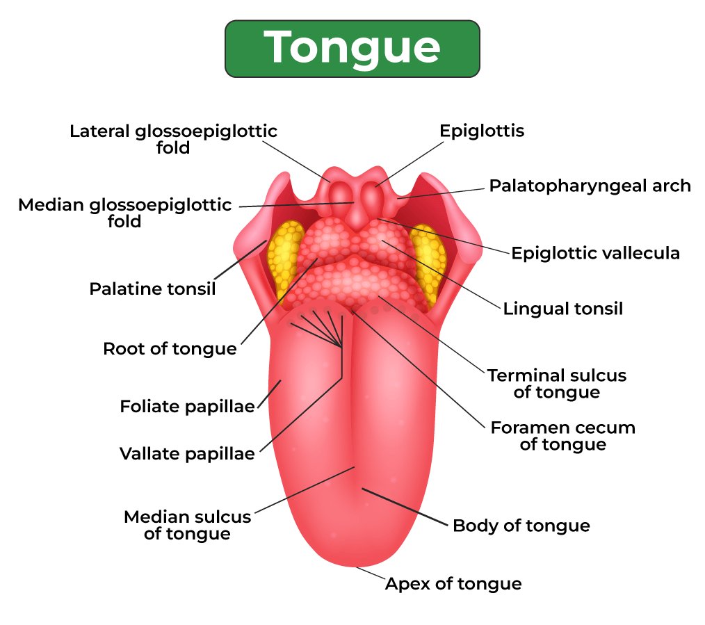
- Tongue: Tongue is an accessory digestive organ composed of skeletal muscle covered with mucous membrane. It helps to taste the food, swallow food and to speak. Tongue is divided into 2 symmetrical lateral parts by a median septum that extends its whole length. Tongue consists of two types of muscles:
- The extrinsic muscles move the tongue from side to side and in and out to maneuver food for chewing, shape the food into a rounded mass, and force the food to the back of the mouth for swallowing. They also form the floor of the mouth and hold the tongue in position.
- The intrinsic muscles alter the shape and size of the tongue for speech and swallowing.
- A fold of mucous membrane in midline of undersurface of tongue called lingual frenulum is attached to floor of mouth and controls posterior movement of tongue. The upper surface and lateral surfaces of the tongue are covered with papillae. Many papillae contain taste buds, the receptors for gustation (taste). Some papillae lack taste buds, but they contain receptors for touch and increase friction between the tongue and food, making it easier for the tongue to move food in the oral cavity. Lingual glands are also present which secretes mucus and fluid containing an enzyme lingual lipase which breakdowns the triglycerides
- Teeth: Humans are diphyodont i.e. they have two sets of teeth- milk or deciduous and permanent teeth. Here is the list of different types of teeth in humans with their functions.
| Type of teeth | Function |
|---|---|
| Incisors | Used for cutting |
| Canines | Used for tearing |
| Premolars | Used for Chopping |
| Molars | Used for Grinding and Chewing |
The structure of the tooth is made up of three parts- the crown, neck, and root. The exposed part of the tooth is called the crown, the region where it is covered with gums is known as neck and root is embedded in the socket of the jaw bone.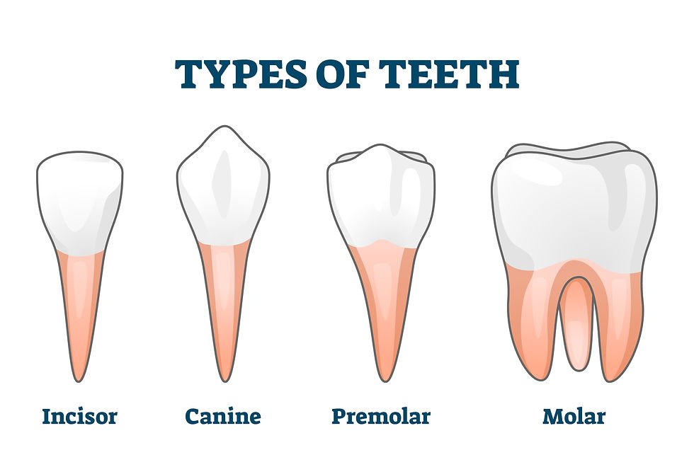
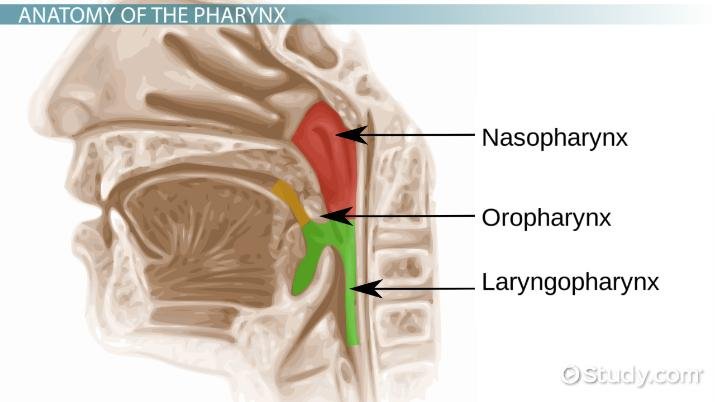
- Pharynx: It is the common passage for food and air. Epiglottis prevents the entry of food into the windpipe. The pharynx delivers food and liquid to your esophagus, which sends them on to your stomach. More than that, your pharynx helps to make sure particles of food and liquid don't tumble into your trachea and your lungs.
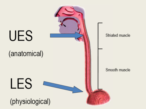
- Oesophagus: The esophagus, historically also spelled oesophagus, is a tubular, elongated organ of the digestive system which connects the pharynx to the stomach. The esophagus is the organ that food travels through to reach the stomach for further digestion. It follows a path that travels behind the trachea and heart, in front of the spinal column, and through the diaphragm before entering the stomach. The organ is typically about nine to ten inches (23 to 25 cm) long in fully grown adults, with sphincters located at each of its proximal and distal extremities, a mucosa-lined lumen and connective tissue, and smooth muscle outer composition. The sphincter located anteriorly, the upper esophageal sphincter, allows for the single direction passage of food into the esophagus, and anteriorly, the lower esophageal sphincter allows for the single direction passage of food into the stomach.
- Stomach: The stomach is a J-shaped organ that digests food. The stomach sits in the upper abdomen on the left side of the body. The top of the stomach connects to a valve called the esophageal sphincter (a muscle at the end of your esophagus). The bottom of the stomach connects to the small intestine. It produces (substances that create chemical reactions) and acids (digestive juices). This mix of enzymes and digestive juices breaks down food so it can pass to the small intestine. The stomach is part of the gastrointestinal (GI) tract. The size of the stomach varies from person to person. It expands when full and deflates when empty. Because of this, the stomach size can vary depending on how recently and how much you have eaten. The GI tract is a key part of the . The stomach’s purpose is to digest food and send it to the small intestine.
It has three functions:
- Temporarily store food.
- Contract and relax to mix and break down food.
- Produce enzymes and other specialized cells to digest food.
The stomach has five distinct sections:
- The cardia is the top part of the stomach. It contains the cardiac sphincter, which prevents food from traveling back up your esophagus.
- The fundus is a rounded section next to the cardia. It's below the diaphragm (the dome-shaped muscle that helps you breathe).
- The body (corpus) is the largest section of the stomach. In the body, the stomach contracts and begins to mix food.
- The antrum lies below the body. It holds food until the stomach is ready to send it to the small intestine.
- The pylorus is the bottom part of the stomach. It includes the pyloric sphincter. This ring of tissue controls when and how the stomach contents move to the small intestine.

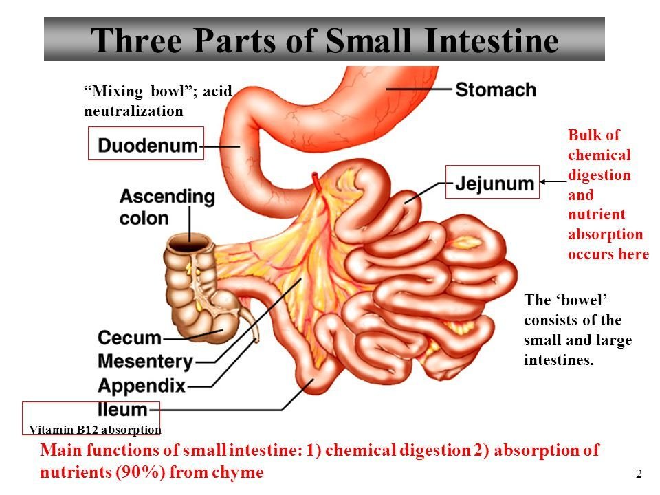
- Small Intestine: The small intestine is the digestive segment spanning from the gastric pylorus' distal tip to the ileocecal valve, the region connecting the small and large bowels. The mesentery is a double peritoneal fold anchoring the jejunum and ileum to the posterior abdominal wall. From the outermost to innermost portion, the small intestinal layers are the serosa, muscularis, submucosa, lamina propria, and mucosa. The serosa is the outermost small intestinal layer, consisting of mesothelium and epithelium. This layer is only present in the jejunum, ileum, and intraperitoneal portions of the duodenum.
The muscularis consists of two smooth muscle layers. The outer layer is longitudinally oriented, while the inner layer is circular. The submucosa consists of a connective tissue layer that contains blood vessels, nerves (sub mucosal or Meissner plexus), and lymphatics.
The small intestine's segments from proximal to distal are the duodenum, jejunum, and ileum. The duodenum is the most proximal, widest, and shortest small bowel section, measuring 20 to 25 cm on average. This portion has superior, descending, inferior, and ascending areas. The first 2 cm of the superior duodenum is mobile, has mesentery, and lies intraperitoneally. The rest of the duodenum is retroperitoneal. This C-shaped small intestine segment curves around the pancreatic head, coursing from the L1 vertebral level 2 to 3 cm left of the midline, descending to L3, and ascending back to L2. The duodenum is the only gut portion where the sub mucosal Brunner glands appear. The jejunum is the small intestine's middle segment, roughly 2.5 meters long. This portion begins at the duodenojejunal junction, where the gastrointestinal tract continues intraperitoneally. The jejunum contains plicae circulares (muscular flaps or valves of Kerckring) and villi. The ileum is the most distal small intestine segment, measuring around 3 meters and terminating at the cecum. Grossly, the jejunum has a deeper red color, thicker wall, and greater vascularity than the ileum. On the other hand, the ileum has more mesenteric fat than the jejunum.
Function
The small intestine's principal function is to break down food, absorb nutrients the body needs, and excrete unnecessary components. This gastrointestinal segment also participates in immune functions, acting as a barrier to intraluminal bacteria. The small bowel also has an endocrinologic role, producing digestive and energy-regulating hormones like cholecystokinin, secretin etc.
Specific small intestinal cells have the following functions:
- Enterocytes are the most abundant cells lining the small intestine's villi. These cells possess microvillus and are specialized for absorbing nutrients.
- Goblet cells are scattered among enterocytes throughout the small bowel's epithelium. These cells secrete mucus, which protects the mucosa from gastrointestinal secretions. Mucus also lubricates food and aids in removing pathogens from the intestinal lumen.
- Paneth cells are found in the crypts of Lieberkühn. These cells secrete antimicrobial peptides like defensins and lysozyme, growth factors, enzymes, and cytokines.
- Enteroendocrine cells are scattered throughout the small intestinal epithelium, particularly in the crypts and villi. These cells secrete various hormones, including cholecystokinin, secretin, gastric inhibitory peptide, and glucagon-like peptide-1.
- Tuft cells (brush cells) are epithelial cells found in the small intestinal villi. These cells have apical microvilli, giving them a "tufted" appearance. Tuft cells sense luminal contents and can secrete various signaling molecules, including cytokines and neurotransmitters, in response to stimuli. Tuft cells help regulate immune responses, mucosal defense, and epithelial repair.
- M cells (microfold cell) are unique epithelial cells in the follicle-associated epithelium covering Peyer patches. These cells function as antigen-sampling cells, transporting luminal antigens and microorganisms across the epithelium to initiate lymphoid immune responses.
- Lymphoid cells aggregate in Peyer patches; the lymph nodules scattered throughout the small intestine but are most abundant in the ileum. These cells are essential to the gut's immune function.
- Stem cells lie within the crypts of Lieberkühn. These multipotent stem cells continuously proliferate and differentiate to replace old or damaged cells. Stem cells give rise to different cell small bowel cell types, including enterocytes and goblet, Paneth, and enteroendocrine cells.
- Taste receptor cells like those on the tongue may be found in the duodenum and jejunum. These cells express taste receptors and detect sweet, umami (savory), and bitter tastes in luminal contents. Small bowel taste receptor cells may play a role in nutrient sensing and gut-brain signaling, influencing food intake and digestion.
Meanwhile, the 3 small intestinal segments have the following functions:
- The duodenum receives chime – a mix of gastric acid and food – from the stomach and is where nutrient absorption begins. Pancreatic enzymes enter the descending segment's posteromedial wall via the hepatopancreatic ampulla, which is regulated by the muscular sphincter of Oddi. Pancreatic enzymes break down chyme. Brunner glands secrete bicarbonate into the duodenum to neutralize stomach acid before chyme reaches the jejunum. The duodenum and upper jejunum are the main dietary iron absorption sites. Bile from the liver also enters the duodenum through the hepatopancreatic ampulla. Bile is essential for lipid digestion and absorption. Smooth muscle contraction helps widen the duodenojejunal junction, assisting in the forward movement of intestinal contents.
- The jejunum primarily absorbs carbohydrates, amino acids, and fatty acids through the villi. Jejunal and ileal plicae circulares increase surface area and enhance nutrient absorption.
- The ileum takes in the nutrients unabsorbed by the first 2 small intestinal components, the most important being vitamin B12 and bile acids. These substances are recycled after absorption.
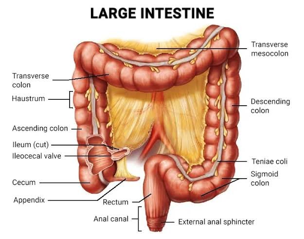
- Large Intestine: It is a long, tube-like organ that is connected to the small intestine at one end and the anus at the other end. The large intestine has four parts: Caecum, colon, rectum, and anal canal. Partly digested food moves through the Caecum into the colon, where water and some nutrients and electrolytes are removed. The large intestine has three primary functions: absorbing water and electrolytes, producing and absorbing vitamins, and forming and propelling feces toward the rectum for elimination.
- Caecum: A pouch that forms the first part of the large intestine. It connects the small intestine to the colon, which is part of the large intestine. The cecum connects the small intestine to the colon. It is a small sac-like structure containing symbiotic microorganisms. The vermiform appendix (vestigial organ) is attached to it.
- Colon: The longest part of the large intestine (a tube-like organ connected to the small intestine at one end and the anus at the other). The colon removes water and some nutrients and electrolytes from partially digested food. It is divided into four regions- ascending, transverse, sigmoid and descending.
- Rectum: The rectum is the lower part of the large intestine that connects to the sigmoid colon. It is about 15 cm (6 in) long. It receives waste from the colon and stores it until it passes out of the body through the anus. It opens into the anus.
- Anal canal: This is the terminal part of the large intestine, measuring about 2.5 to 4 centimeters in length. It extends from the rectum to the anus and plays a crucial role in the control of defecation. The canal is lined with different types of epithelium: the upper part with simple columnar epithelium similar to the rectum, transitioning to stratified squamous epithelium in the lower part near the anus.
Anatomically, the anal canal is divided into two zones:
- Upper Zone (Anal Columns): This zone contains longitudinal folds called anal columns, which are connected by anal valves and form small recesses called anal sinuses. These structures help in maintaining continence and the passage of stool.
- Lower Zone: This is the distal part of the canal, lined by stratified squamous epithelium, which becomes keratinized near the external opening.
The anal canal is surrounded by two sphincters:
- Internal Anal Sphincter: An involuntary muscle that maintains baseline tone to prevent the passage of feces.
- External Anal Sphincter: A voluntary muscle that provides additional control over defecation.
The canal also contains an extensive nerve supply, allowing for precise control and sensation, which is essential for continence and the detection of rectal contents.